
Article Figures & Data
Figures
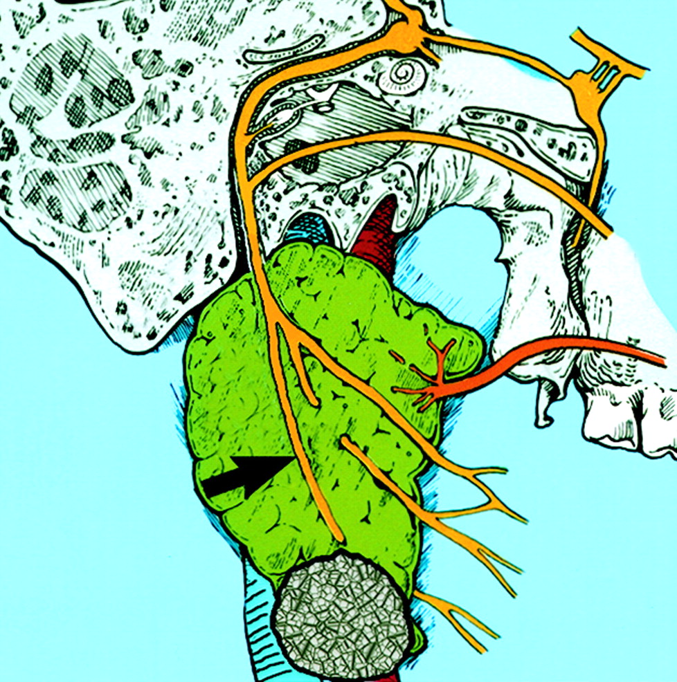
- Fig 1.
Diagram of parotid gland with a typical mass in the tail. Course of the facial nerve is demonstrated. Note the relationship of the marginal mandibular nerve (arrow) as it traverses the parotid tail mass. (Reprinted with permission from reference 1.)
- Fig 2.
Axial contrast-enhanced CT scan demonstrates normal parotid tail anatomy. Anatomic boundaries of the parotid tail are the following: lateral and superficial boundary, platysma (white arrow); anteromedial boundary, posterior belly of digastric (black arrow); posteromedial boundary, SCM (curved white arrow). (Reprinted with permission from reference 1.)
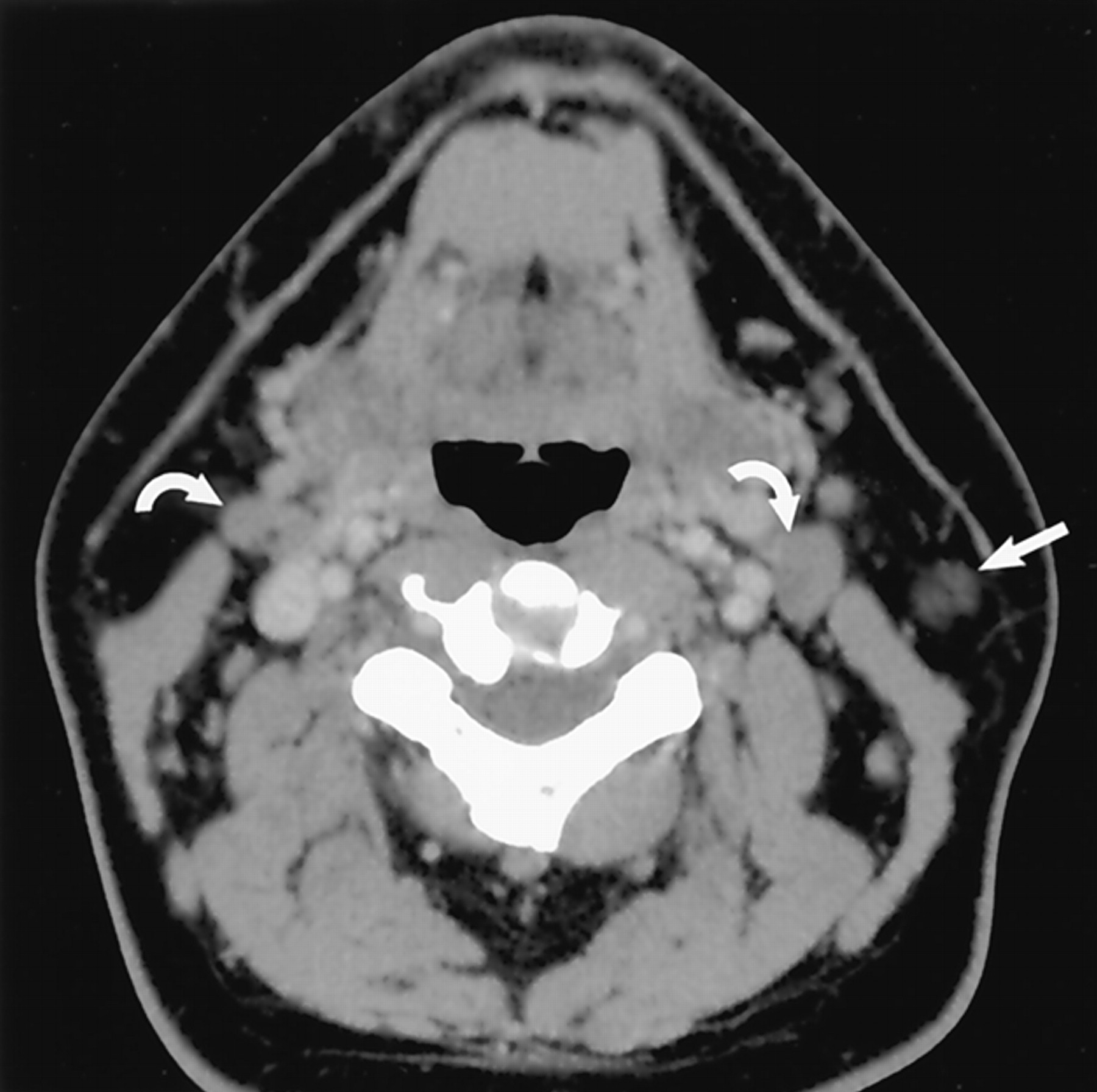
- Fig 3.
Axial contrast-enhanced CT scan demonstrates normal jugulodigastric lymph nodes bilaterally (curved arrows), which should not be confused with lesions originating in the parotid tail. Note the normal parotid tail tissue located anterolateral to the SCM (straight arrow). The lymph nodes lie anteromedial to the muscle. (Reprinted with permission from reference 1.)
- Fig 4.
Earring lesion of the parotid tail. Axial contrast-enhanced CT scan shows typical appearance of a cystic Warthin tumor. Note the nodule of soft tissue (straight white arrow), which is commonly seen. A small amount of parotid tissue (open arrow) is seen anterior to the mass. Note parotid tail anatomic landmarks: SCM posteriorly and platysma (curved white arrow) laterally. (Reprinted with permission from reference 1.)
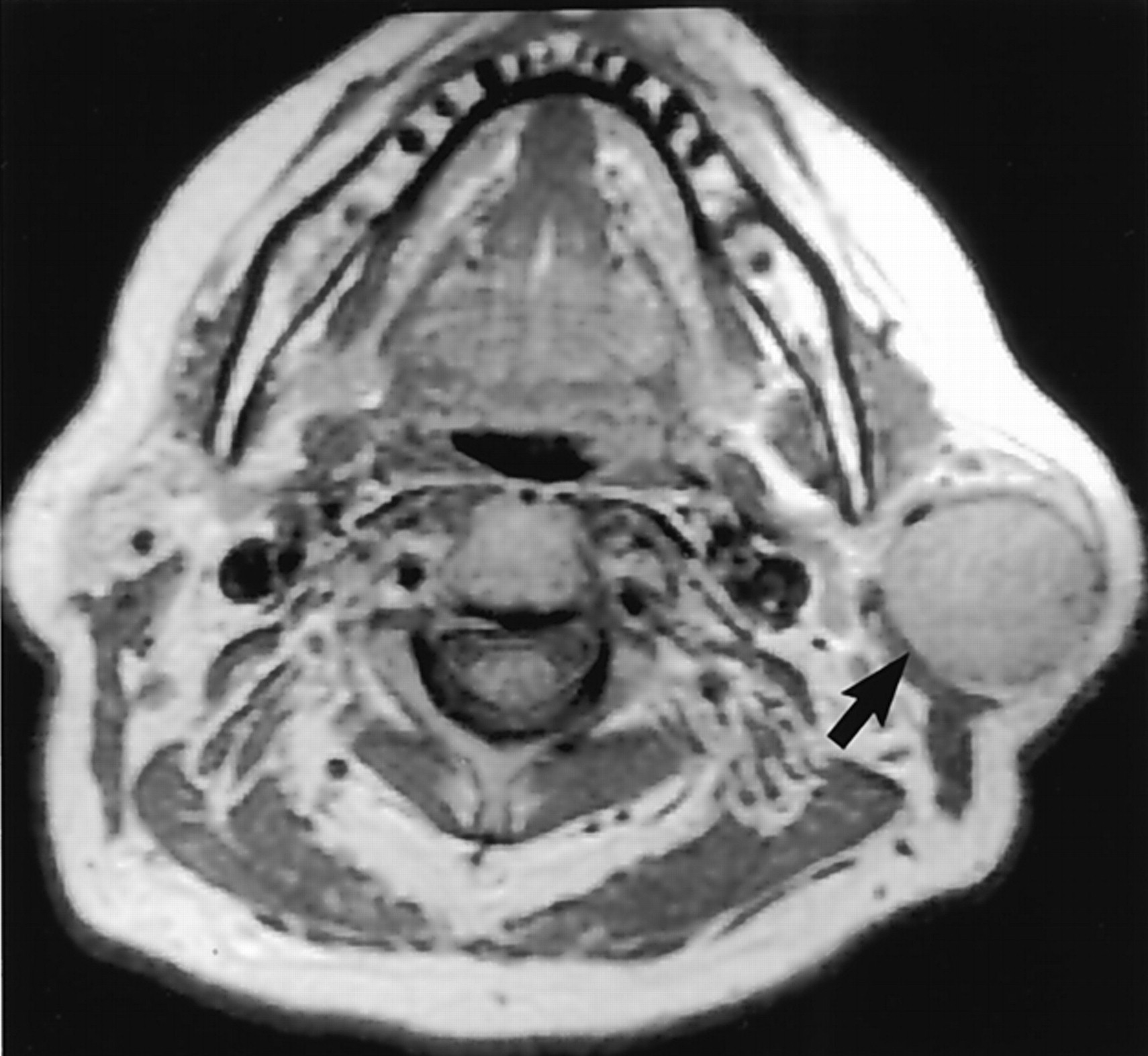
- Fig 5.
Axial nonenhanced T1-weighted MR image demonstrates a large mass (arrow) in the left parotid tail. This mass has little inherent contrast against adjacent normal tissues, making localization more challenging. (Reprinted with permission from reference 1.)
- Fig 6.
Coronal nonenhanced T1-weighted MR image clarifies the intraparotid nature of this BMT (arrow). (Reprinted with permission from reference 1.)
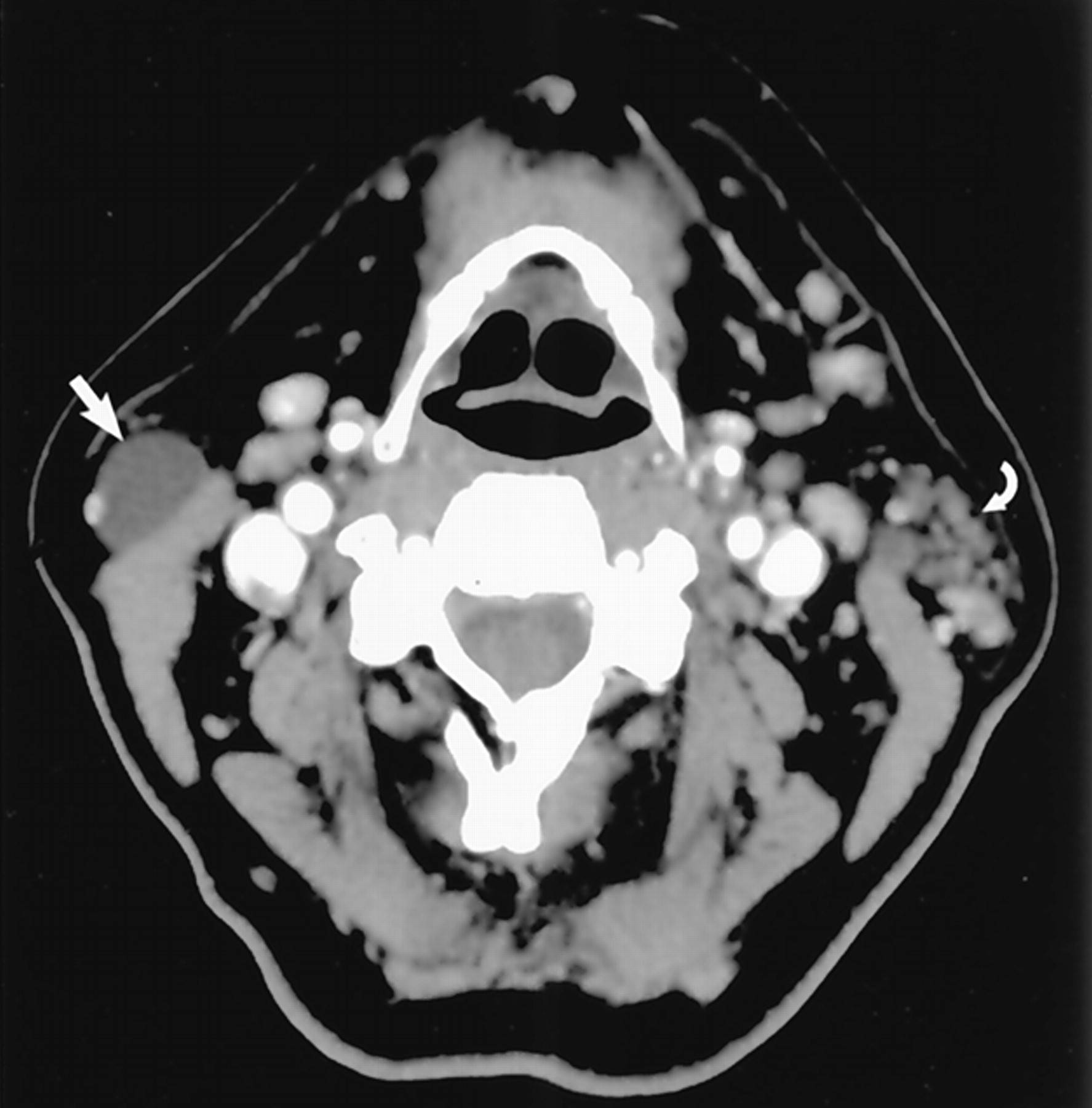
- Fig 7.
Mass was believed to represent a submandibular space or gland tumor by the referring general surgeon, who initially planned to excise the mass. Axial contrast-enhanced CT image reveals the mass (straight arrow) to be deep to platysma and anterior to the SCM, characteristic for a parotid tail “earring lesion.” Note its proximity to the submandibular gland (curved arrow). (Reprinted with permission from reference 1.)
- Fig 8.
Otolaryngology referred this patient for imaging after palpating a mass in the “left parotid tail.” Axial contrast-enhanced CT scan through the mass reveals a partly cystic mass in a typical location for a jugulodigastric lymph node (curved arrow). Normal parotid tail tissue is seen (straight white arrow). This was a metastatic node from a clinically occult primary squamous cell carcinoma, which is partly seen on this section in the left faucial tonsil (open arrow). Additional lymph nodes lower in the neck indicated a high-stage tumor. Upon chart review, the referring physician had noted mass effect in the left tonsil; however, a mucosal lesion was not present. (Reprinted with permission from reference 1.)
- Fig 9.
Axial contrast-enhanced CT scan through the parotid tail demonstrates a dominant cyst (straight arrow). Nodular lymphoid aggregates (curved arrow) in the contralateral gland should suggest a systemic process, in this case, Sjögren disease. The appearance is typical of Sjögren disease or HIV lymphoepithelial lesion, which cannot be distinguished on a histopathologic basis. (Reprinted with permission from reference 1.)


Tables
Distribution of 111 parotid tail masses in 103 patients
Lesion Number Benign (n = 84) Pleomorphic adenoma (BMT) 15 Warthin tumor 14 Infectious process 13 Venous malformation 9 Sjögren disease 9 Lymphatic malformation 7 Lipoma 6 HIV lymphoepithelial lesion 4 First branchial cleft cyst 3 Oncocytoma 2 Sarcoid 1 Lymph node 1 Malignant (n = 27) Non-Hodgkin lymphoma 14 Metastatic disease 7 Mucoepidermoid carcinoma 4 Acinic cell carcinoma 1 Undifferentiated carcinoma 1 Note.—Eight patients had two diagnoses (see text). BMT indicates benign mixed tumors; HIV, human immunodeficiency virus.
In this issue


Jump to section
Related Articles
Cited By...
- No citing articles found.