Advertisement

Main menu

  • Home
  • Content
    • Current Issue
    • Accepted Manuscripts
    • Article Preview
    • Past Issue Archive
    • Video Articles
    • AJNR Case Collection
    • Case of the Week Archive
    • Case of the Month Archive
    • Classic Case Archive
  • Special Collections
    • AJNR Awards
    • Low-Field MRI
    • Alzheimer Disease
    • ASNR Foundation Special Collection
    • Photon-Counting CT
    • View All
  • Multimedia
    • AJNR Podcasts
    • AJNR SCANtastic
    • Trainee Corner
    • MRI Safety Corner
    • Imaging Protocols
  • For Authors
    • Submit a Manuscript
    • Submit a Video Article
    • Submit an eLetter to the Editor/Response
    • Manuscript Submission Guidelines
    • Statistical Tips
    • Fast Publishing of Accepted Manuscripts
    • Graphical Abstract Preparation
    • Imaging Protocol Submission
    • Author Policies
  • About Us
    • About AJNR
    • Editorial Board
    • Editorial Board Alumni
  • More
    • Become a Reviewer/Academy of Reviewers
    • Subscribers
    • Permissions
    • Alerts
    • Feedback
    • Advertisers
    • ASNR Home

User menu

  • Alerts
  • Log in

Search

  • Advanced search
American Journal of Neuroradiology

American Journal of Neuroradiology

ASHNR American Society of Functional Neuroradiology ASHNR American Society of Pediatric Neuroradiology ASSR
  • Alerts
  • Log in

Advanced Search

  • Home
  • Content
    • Current Issue
    • Accepted Manuscripts
    • Article Preview
    • Past Issue Archive
    • Video Articles
    • AJNR Case Collection
    • Case of the Week Archive
    • Case of the Month Archive
    • Classic Case Archive
  • Special Collections
    • AJNR Awards
    • Low-Field MRI
    • Alzheimer Disease
    • ASNR Foundation Special Collection
    • Photon-Counting CT
    • View All
  • Multimedia
    • AJNR Podcasts
    • AJNR SCANtastic
    • Trainee Corner
    • MRI Safety Corner
    • Imaging Protocols
  • For Authors
    • Submit a Manuscript
    • Submit a Video Article
    • Submit an eLetter to the Editor/Response
    • Manuscript Submission Guidelines
    • Statistical Tips
    • Fast Publishing of Accepted Manuscripts
    • Graphical Abstract Preparation
    • Imaging Protocol Submission
    • Author Policies
  • About Us
    • About AJNR
    • Editorial Board
    • Editorial Board Alumni
  • More
    • Become a Reviewer/Academy of Reviewers
    • Subscribers
    • Permissions
    • Alerts
    • Feedback
    • Advertisers
    • ASNR Home
  • Follow AJNR on Twitter
  • Visit AJNR on Facebook
  • Follow AJNR on Instagram
  • Join AJNR on LinkedIn
  • RSS Feeds

AJNR Awards, New Junior Editors, and more. Read the latest AJNR updates

Research ArticleBrain

Thalamic Involvement in Sporadic Creutzfeldt-Jakob Disease: A Diffusion-Weighted MR Imaging Study

Henriette J. Tschampa, Petra Mürtz, Sebastian Flacke, Sebastian Paus, Hans H. Schild and Horst Urbach
American Journal of Neuroradiology May 2003, 24 (5) 908-915;
Henriette J. Tschampa
  • Find this author on Google Scholar
  • Find this author on PubMed
  • Search for this author on this site
Petra Mürtz
  • Find this author on Google Scholar
  • Find this author on PubMed
  • Search for this author on this site
Sebastian Flacke
  • Find this author on Google Scholar
  • Find this author on PubMed
  • Search for this author on this site
Sebastian Paus
  • Find this author on Google Scholar
  • Find this author on PubMed
  • Search for this author on this site
Hans H. Schild
  • Find this author on Google Scholar
  • Find this author on PubMed
  • Search for this author on this site
Horst Urbach
  • Find this author on Google Scholar
  • Find this author on PubMed
  • Search for this author on this site
  • Article
  • Figures & Data
  • Info & Metrics
  • Responses
  • References
  • PDF
Loading

Article Figures & Data

Figures

  • Tables
  • Fig 1.
    • Download figure
    • Open in new tab
    • Download powerpoint
    Fig 1.

    Patient 5. DW MR images (b = 1000 s/mm2) (A and B) and corresponding ADC maps (C and D) show the level of the basal ganglia (A and C) and the hippocampus (B and D). The marked areas show the region-of-interest measurements in the head of the caudate nucleus, putamen, pulvinar thalami, and MD (A and C) and in the hippocampus (B and D).
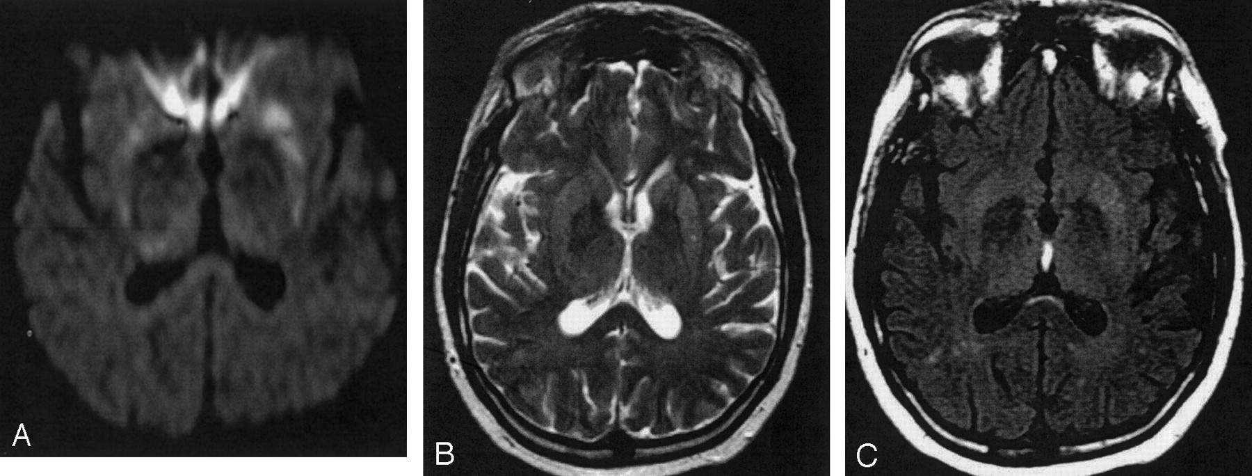
  • Fig 2.
    • Download figure
    • Open in new tab
    • Download powerpoint
    Fig 2.

    Patient 1, second examination. DW image (A), T2-weighted TSE (B), and FLAIR (C) images. Hyperintense SI changes in the striatum and the pulvinar thalami are discernable on the DW (A) and FLAIR (C) images bilaterally, whereas on the T2-weighted TSE image, increased SI is discernable only in the striatum bilaterally.

  • Fig 3.
    • Download figure
    • Open in new tab
    • Download powerpoint
    Fig 3.

    Mean ADCs (± SEM) of sCJD patients (right dark columns) and controls (left bright columns). ADCs in sCJD patients are significantly reduced in the caudate nucleus, putamen, MD, and pulvinar thalami as compared with controls. In the hippocampus (last pair of columns on the right), ADCs are comparable.

  • Fig 4.
    • Download figure
    • Open in new tab
    • Download powerpoint
    Fig 4.

    Patient 1. Serial DW images (A–C) and corresponding ADC maps (D–F) acquired 3 (A and D), 4 (B and E), and 8 (C and F) months after onset of symptoms. The DW image acquired 3 months after onset of symptoms (A) shows bilateral high SI in the caudate nucleus and the rostral part of the putamen. Four months after onset of symptoms, the DW image (B) shows high SI also in the dorsal part of the putamen and additionally in the pulvinar thalami. Eight months after onset of symptoms, the DW image (C) shows severe brain atrophy with widening of the ventricles and of the sulci. The head of the caudate nucleus on the left side totally disappeared; the right caudate nucleus and the putamen on both sides show severe atrophy. There is high SI in the right caudate nucleus and putamen bilaterally and in the frontal, parietal, and cingulate cortex. The ADC maps (D–F) show low SI in the hyperintense areas on DW images corresponding to restricted diffusion.
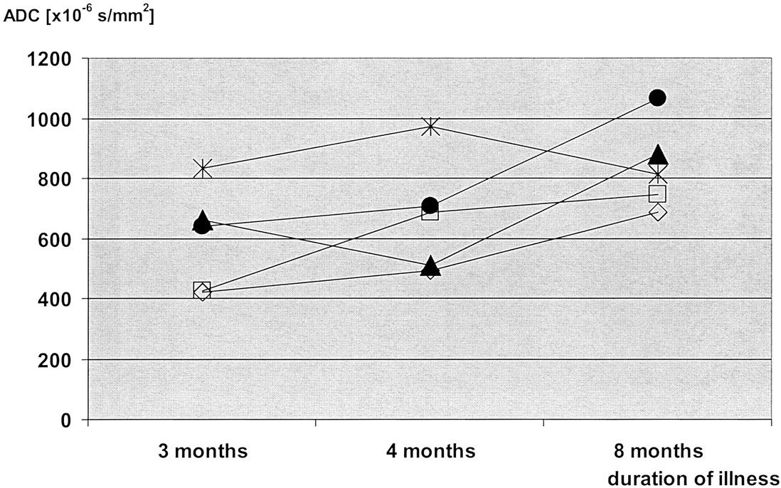
  • Fig 5.
    • Download figure
    • Open in new tab
    • Download powerpoint
    Fig 5.

    Patient 1. Time courses of the ADC values in serial images. Measurements were performed in five anatomic regions: the head of the caudate nucleus (empty rectangle), the putamen (open rhomboid), the pulvinar thalami (filled triangle), the MD (filled circle), and the hippocampus (star). In all areas except for the hippocampus, ADCs were lowest in the first examination and highest in the last examination. In the caudate nucleus, the putamen, and the MD, there was a gradual increase in the ADCs during the course of the disease. In the pulvinar thalami, ADCs were lowest in the second examination; this was paralleled by an increased SI on DW images in the second examination (shown in Fig 4B, E).

Tables

  • Figures
    • View popup
    TABLE 1:

    Clinical data of sporadic CJD patients

    Patient
    123456
    Age at onset56.2 years58.9 years71.9 years62.9 years63.6 years61 years
    GenderMaleMaleMaleFemaleMaleFemale
    Symptoms/signsDementia, ataxia, myoclonusDementia, vertigo, ataxiaDementia, ataxia, myoclonusDementia, vertigo, gait disturbancesDementia, ataxia, signs of pyramidal tractDementia, vertigo, ataxia
    EEGNot typical for CJDNot typical for CJDPSWCsaPSWCsaPSWCsNot typical for CJD
    Protein 14-3-3PositivePositivePositivePositivePositivePositive
    Diagnosis of CJDbProbableProbableProbableProbableDefiniteProbable
    • a PSWCs are periodic sharp wave complexes (4).

    • b WHO 1998 (1).

    • View popup
    TABLE 2:

    Analysis of DW-, T2-, and FLAIR-weighted images

    PulvinarMDaPutamenCaudate NucleusHippocampusOther
    Patient 1:b
     DW images——↑ SI bilat.↑ SI bilat.——
     T2w images——↑ SI bilat.↑ SI l. > r.——
     FLAIR images——↑ SI bilat.↑ SI bilat.
    Patient 2:
     DW images(↑ SI bilat.)—↑ SI bilat.↑ SI bilat.——
     T2w images——↑ SI l.↑ SI l.——
     FLAIR images(↑ SI bilat.)—↑ SI bilat.↑ SI bilat.—
    Patient 3:
     DW images——(↑ SI bilat.)(↑ SI bilat.)——
     T2w images——(↑ SI bilat.)——
     FLAIR images——(↑ SI bilat.)(↑ SI bilat.)—
    Patient 4:
     DW images↑ SI bilat.—↑ SI bilat.↑ SI bilat.——
     T2w images——————
     FLAIR images (coronal)—d—d↑ SI bilat.↑ SI bilat.——
    Patient 5:
     DW images↑ SI bilat.—↑ SI bilat.↑ SI bilat.—↑ SI Cerebellar cortex
     b0 images of DW imagingc——↑ SI r.↑ SI r.——
     FLAIR images——↑ SI bilat.↑ SI bilat.—↑ SI Cerebellar cortex
    Patient 6:
     DW images↑ SI bilat.—↑ SI bilat.↑ SI bilat.——
     T2w images——↑ SI bilat.↑ SI bilat.——
     FLAIR images↑ SI bilat.—↑ SI bilat.↑ SI bilat.——
    • Note.—Parentheses indicate slight change. An upward-pointing arrow (↑) indicates that a value has increased. A dash (—) indicates no signal change; bilat., bilateral; l., left; r., right; SI, signal intensity.

    • a Mediodorsal thalamic nucleus.

    • b The first of the three consecutive measurements of patient 1 is considered.

    • c b0 images of the DW imaging scan were used, because no T2-weighted TSE images were acquired.

    • d Disturbed by motion artifacts.

    • View popup
    TABLE 3:

    ADC values [× 10−6 s/mm2; b = 1000 s/mm2] in sCJD patients and controls

    AreaPatientsa (mean ± SEM)Controls (mean ± SEM)Pb (unpaired two-tailed Student t test)
    Caudate nucleus548 ± 27767 ± 35<0.0001
    Putamen585 ± 28754 ± 22<0.0001
    Mediodorsal thalamic nucleus664 ± 28800 ± 240.0011
    Pulvinar:
     1. All patients695 ± 27853 ± 15<0.0001
     2. Patients with SI changes701 ± 38c
     3. Patients without SI changes684 ± 37
    Hippocampus853 ± 22871 ± 210.56
    • a In patient 1 in Table 1, the first of the three consecutive measurements was considered.

    • b P < .05 is considered significant.

    • c P = .78 (not significant) comparing sCJD patients with and those without SI changes in the thalamus.

PreviousNext
Back to top

In this issue

American Journal of Neuroradiology: 24 (5)
American Journal of Neuroradiology
Vol. 24, Issue 5
1 May 2003
  • Table of Contents
  • Index by author
Advertisement
Print
Download PDF
Email Article

Thank you for your interest in spreading the word on American Journal of Neuroradiology.

NOTE: We only request your email address so that the person you are recommending the page to knows that you wanted them to see it, and that it is not junk mail. We do not capture any email address.

Enter multiple addresses on separate lines or separate them with commas.
Thalamic Involvement in Sporadic Creutzfeldt-Jakob Disease: A Diffusion-Weighted MR Imaging Study
(Your Name) has sent you a message from American Journal of Neuroradiology
(Your Name) thought you would like to see the American Journal of Neuroradiology web site.
CAPTCHA
This question is for testing whether or not you are a human visitor and to prevent automated spam submissions.
Cite this article
Henriette J. Tschampa, Petra Mürtz, Sebastian Flacke, Sebastian Paus, Hans H. Schild, Horst Urbach
Thalamic Involvement in Sporadic Creutzfeldt-Jakob Disease: A Diffusion-Weighted MR Imaging Study
American Journal of Neuroradiology May 2003, 24 (5) 908-915;

Citation Manager Formats

  • BibTeX
  • Bookends
  • EasyBib
  • EndNote (tagged)
  • EndNote 8 (xml)
  • Medlars
  • Mendeley
  • Papers
  • RefWorks Tagged
  • Ref Manager
  • RIS
  • Zotero
0 Responses
Respond to this article
Share
Bookmark this article
Thalamic Involvement in Sporadic Creutzfeldt-Jakob Disease: A Diffusion-Weighted MR Imaging Study
Henriette J. Tschampa, Petra Mürtz, Sebastian Flacke, Sebastian Paus, Hans H. Schild, Horst Urbach
American Journal of Neuroradiology May 2003, 24 (5) 908-915;
del.icio.us logo Twitter logo Facebook logo Mendeley logo
  • Tweet Widget
  • Facebook Like
  • Google Plus One

Jump to section

  • Article
    • Abstract
    • Methods
    • Results
    • Discussion
    • Conclusion
    • References
  • Figures & Data
  • Info & Metrics
  • Responses
  • References
  • PDF

Related Articles

  • No related articles found.
  • PubMed
  • Google Scholar

Cited By...

  • Mediodorsal nucleus and its multiple cognitive functions
  • High-b-Value Diffusion MR Imaging and Basal Nuclei Apparent Diffusion Coefficient Measurements in Variant and Sporadic Creutzfeldt-Jakob Disease
  • Brain-water diffusion coefficients reflect the severity of inherited prion disease
  • Enhanced Detection of Diffusion Reductions in Creutzfeldt-Jakob Disease at a Higher B Factor
  • Pathologic correlates of diffusion MRI changes in Creutzfeldt-Jakob disease
  • Neuroimaging findings in human prion disease
  • Crossref
  • Google Scholar

This article has not yet been cited by articles in journals that are participating in Crossref Cited-by Linking.

More in this TOC Section

  • Progression of Microstructural Damage in Spinocerebellar Ataxia Type 2: A Longitudinal DTI Study
  • SWI or T2*: Which MRI Sequence to Use in the Detection of Cerebral Microbleeds? The Karolinska Imaging Dementia Study
  • Statin Therapy Does Not Affect the Radiographic and Clinical Profile of Patients with TIA and Minor Stroke
Show more Brain

Similar Articles

Advertisement

Indexed Content

  • Current Issue
  • Accepted Manuscripts
  • Article Preview
  • Past Issues
  • Editorials
  • Editor's Choice
  • Fellows' Journal Club
  • Letters to the Editor
  • Video Articles

Cases

  • Case Collection
  • Archive - Case of the Week
  • Archive - Case of the Month
  • Archive - Classic Case

More from AJNR

  • Trainee Corner
  • Imaging Protocols
  • MRI Safety Corner
  • Book Reviews

Multimedia

  • AJNR Podcasts
  • AJNR Scantastics

Resources

  • Turnaround Time
  • Submit a Manuscript
  • Submit a Video Article
  • Submit an eLetter to the Editor/Response
  • Manuscript Submission Guidelines
  • Statistical Tips
  • Fast Publishing of Accepted Manuscripts
  • Graphical Abstract Preparation
  • Imaging Protocol Submission
  • Evidence-Based Medicine Level Guide
  • Publishing Checklists
  • Author Policies
  • Become a Reviewer/Academy of Reviewers
  • News and Updates

About Us

  • About AJNR
  • Editorial Board
  • Editorial Board Alumni
  • Alerts
  • Permissions
  • Not an AJNR Subscriber? Join Now
  • Advertise with Us
  • Librarian Resources
  • Feedback
  • Terms and Conditions
  • AJNR Editorial Board Alumni

American Society of Neuroradiology

  • Not an ASNR Member? Join Now

© 2025 by the American Society of Neuroradiology All rights, including for text and data mining, AI training, and similar technologies, are reserved.
Print ISSN: 0195-6108 Online ISSN: 1936-959X

Powered by HighWire