Advertisement

Main menu

  • Home
  • Content
    • Current Issue
    • Accepted Manuscripts
    • Article Preview
    • Past Issue Archive
    • Video Articles
    • AJNR Case Collection
    • Case of the Week Archive
    • Case of the Month Archive
    • Classic Case Archive
  • Special Collections
    • AJNR Awards
    • Low-Field MRI
    • Alzheimer Disease
    • ASNR Foundation Special Collection
    • Photon-Counting CT
    • View All
  • Multimedia
    • AJNR Podcasts
    • AJNR SCANtastic
    • Trainee Corner
    • MRI Safety Corner
    • Imaging Protocols
  • For Authors
    • Submit a Manuscript
    • Submit a Video Article
    • Submit an eLetter to the Editor/Response
    • Manuscript Submission Guidelines
    • Statistical Tips
    • Fast Publishing of Accepted Manuscripts
    • Graphical Abstract Preparation
    • Imaging Protocol Submission
    • Author Policies
  • About Us
    • About AJNR
    • Editorial Board
    • Editorial Board Alumni
  • More
    • Become a Reviewer/Academy of Reviewers
    • Subscribers
    • Permissions
    • Alerts
    • Feedback
    • Advertisers
    • ASNR Home

User menu

  • Alerts
  • Log in

Search

  • Advanced search
American Journal of Neuroradiology

American Journal of Neuroradiology

ASHNR American Society of Functional Neuroradiology ASHNR American Society of Pediatric Neuroradiology ASSR
  • Alerts
  • Log in

Advanced Search

  • Home
  • Content
    • Current Issue
    • Accepted Manuscripts
    • Article Preview
    • Past Issue Archive
    • Video Articles
    • AJNR Case Collection
    • Case of the Week Archive
    • Case of the Month Archive
    • Classic Case Archive
  • Special Collections
    • AJNR Awards
    • Low-Field MRI
    • Alzheimer Disease
    • ASNR Foundation Special Collection
    • Photon-Counting CT
    • View All
  • Multimedia
    • AJNR Podcasts
    • AJNR SCANtastic
    • Trainee Corner
    • MRI Safety Corner
    • Imaging Protocols
  • For Authors
    • Submit a Manuscript
    • Submit a Video Article
    • Submit an eLetter to the Editor/Response
    • Manuscript Submission Guidelines
    • Statistical Tips
    • Fast Publishing of Accepted Manuscripts
    • Graphical Abstract Preparation
    • Imaging Protocol Submission
    • Author Policies
  • About Us
    • About AJNR
    • Editorial Board
    • Editorial Board Alumni
  • More
    • Become a Reviewer/Academy of Reviewers
    • Subscribers
    • Permissions
    • Alerts
    • Feedback
    • Advertisers
    • ASNR Home
  • Follow AJNR on Twitter
  • Visit AJNR on Facebook
  • Follow AJNR on Instagram
  • Join AJNR on LinkedIn
  • RSS Feeds

AJNR Awards, New Junior Editors, and more. Read the latest AJNR updates

Research ArticleNeurointervention

Guglielmi Detachable Coil Treatment of Ruptured Saccular Cerebral Aneurysms: Retrospective Review of a 10-Year Single-Center Experience

Jonathan A. Friedman, Douglas A. Nichols, Fredric B. Meyer, Mark A. Pichelmann, Jon I. McIver, L. Gerard Toussaint, Patsy L. Axley and Robert D. Brown
American Journal of Neuroradiology March 2003, 24 (3) 526-533;
Jonathan A. Friedman
  • Find this author on Google Scholar
  • Find this author on PubMed
  • Search for this author on this site
Douglas A. Nichols
  • Find this author on Google Scholar
  • Find this author on PubMed
  • Search for this author on this site
Fredric B. Meyer
  • Find this author on Google Scholar
  • Find this author on PubMed
  • Search for this author on this site
Mark A. Pichelmann
  • Find this author on Google Scholar
  • Find this author on PubMed
  • Search for this author on this site
Jon I. McIver
  • Find this author on Google Scholar
  • Find this author on PubMed
  • Search for this author on this site
L. Gerard Toussaint III
  • Find this author on Google Scholar
  • Find this author on PubMed
  • Search for this author on this site
Patsy L. Axley
  • Find this author on Google Scholar
  • Find this author on PubMed
  • Search for this author on this site
Robert D. Brown Jr
  • Find this author on Google Scholar
  • Find this author on PubMed
  • Search for this author on this site
  • Article
  • Figures & Data
  • Info & Metrics
  • Responses
  • References
  • PDF
Loading

Article Figures & Data

Figures

  • Tables
  • Fig 1.
    • Download figure
    • Open in new tab
    • Download powerpoint
    Fig 1.

    Graph depicting patients with ruptured cerebral aneurysms treated with endovascular GDC occlusion by year.

  • Fig 2.
    • Download figure
    • Open in new tab
    • Download powerpoint
    Fig 2.

    A 56-year-old man with Hunt-Hess grade 1 subarachnoid hemorrhage.

    A, Towne-view right vertebral DSA demonstrates an 8-mm basilar caput aneurysm. The aneurysm was treated with seven GDCs measuring a total length of 51 cm.

    B, Immediate post-treatment Towne-view right vertebral DSA demonstrates total occlusion of the aneurysm. Follow-up angiography obtained 6 months after treatment demonstrated persistent total occlusion of the aneurysm (not shown).

  • Fig 3.
    • Download figure
    • Open in new tab
    • Download powerpoint
    Fig 3.

    A 39-year-old man with Hunt-Hess grade 1 subarachnoid hemorrhage.

    A, Towne-view left vertebral DSA demonstrates a 16-mm basilar caput aneurysm. The aneurysm was treated with seven GDCs measuring a total length of 180 cm. Immediate post-treatment DSA demonstrated a small “dog ear” neck remnant (not shown).

    B, Towne-view left vertebral DSA obtained 6 weeks post treatment demonstrates slight enlargement of the “dog ear” neck remnant (arrow) at the right base of the aneurysm.

    C, Towne-view left vertebral DSA obtained immediately after detachment of one GDC measuring 8 cm in the dog ear neck remnant demonstrates occlusion of the neck remnant and total occlusion of the aneurysm.

  • Fig 4.
    • Download figure
    • Open in new tab
    • Download powerpoint
    Fig 4.

    A 88-year-old man with Hunt-Hess grade 2 subarachnoid hemorrhage.

    A, Lateral view right internal carotid DSA demonstrates a 3 × 6-mm anterior choroidal artery aneurysm. Oblique views (not shown) demonstrated direct origin of the anterior choroidal artery from the neck of the aneurysm. Significant atherosclerotic disease involving the cavernous right internal carotid artery and the right middle cerebral artery is also demonstrated.

    B, Lateral view right internal carotid DSA immediately after detachment of four GDCs measuring a total length of 20 cm demonstrate occlusion of the fundus and dome of the aneurysm. A residual neck remnant (arrow) was intentionally left to preserve the origin of the anterior choroidal artery.
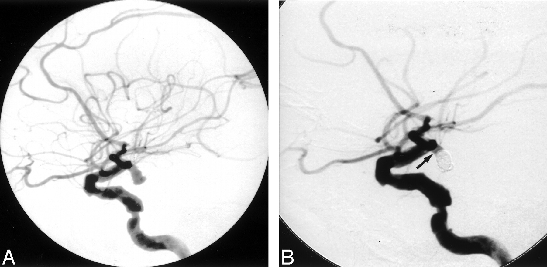
  • Fig 5.
    • Download figure
    • Open in new tab
    • Download powerpoint
    Fig 5.

    A 66-year-old woman with Hunt-Hess grade 3 subarachnoid hemorrhage.

    A, Lateral view left internal carotid DSA demonstrates a 15-mm superior hypophyseal aneurysm. Seven GDCs measuring a total length of 140 cm were detached in the aneurysm. Immediate post-treatment DSA demonstrated persistent opacification of the aneurysm.

    B, Six-month follow-up lateral view left internal carotid DSA demonstrates a residual aneurysm. The aneurysm was subsequently surgically clipped without complications.
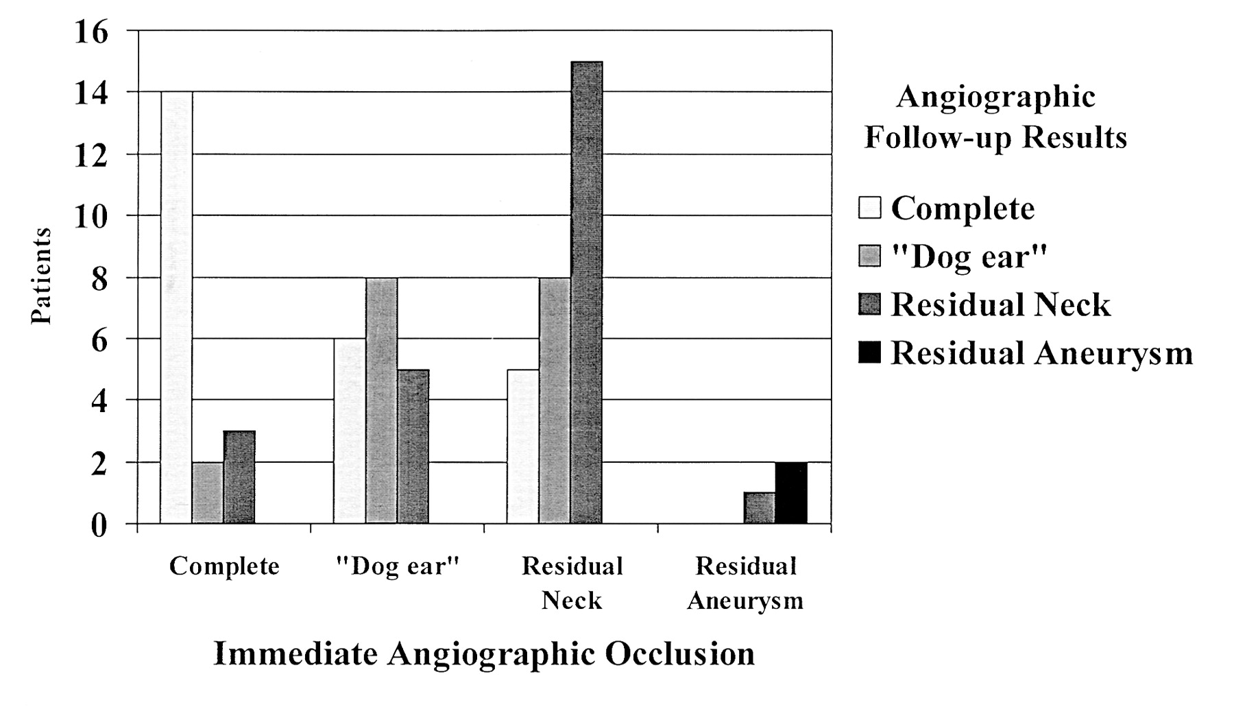
  • Fig 6.
    • Download figure
    • Open in new tab
    • Download powerpoint
    Fig 6.

    Graph depicting degree of occlusion at follow-up angiography with respect to initial angiographic result.
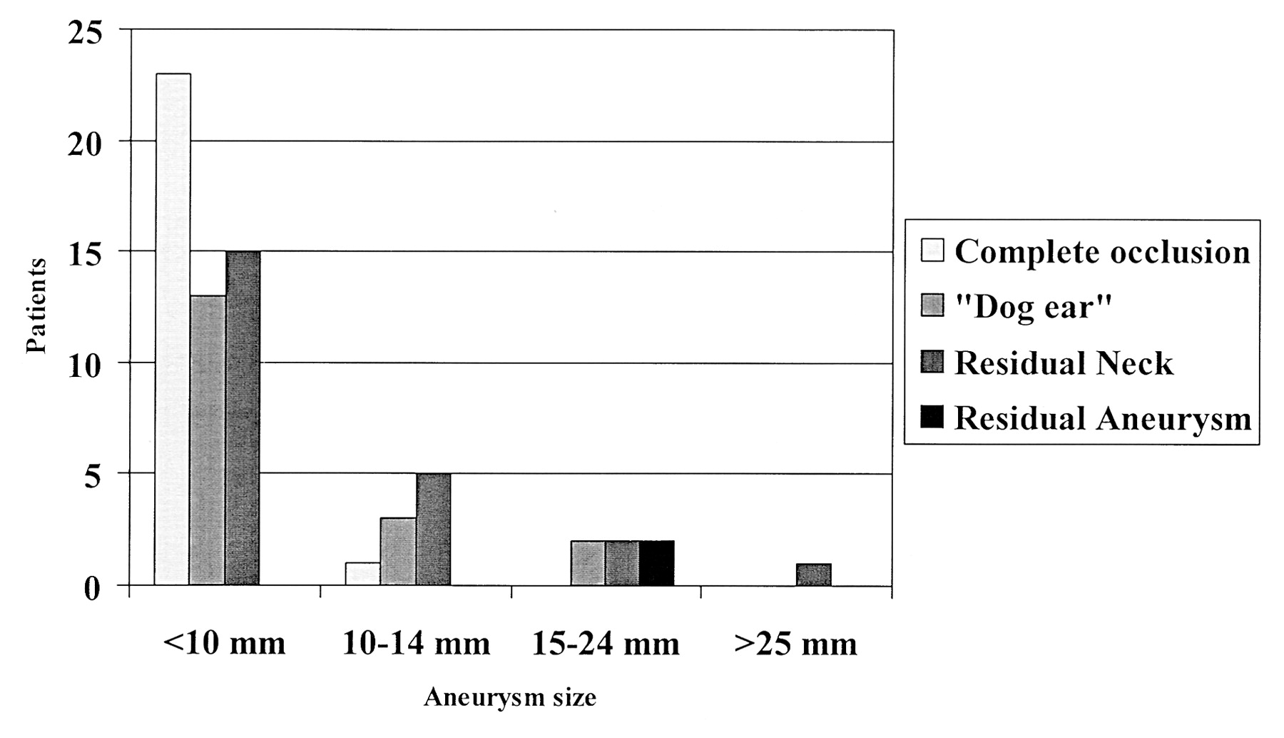
  • Fig 7.
    • Download figure
    • Open in new tab
    • Download powerpoint
    Fig 7.

    Graph depicting angiographic outcome at longest angiographic follow-up with respect to initial aneurysm size.

Tables

  • Figures
    • View popup
    TABLE 1:

    Patient data

    CharacteristicValue
    Sex
     Male33
     Female50
    Age, y
     Mean56.1
     Range27–88
    Hunt-Hess grade at admission
     I33
     II14
     III20
     IV12
     V3
     Mean2.2
    Clinical follow-up, mo
     Mean16.8
     Range0.25–112
     Mean in survivors19.1
     Range in survivors0.5–112
    Patients with angiographic follow-up68
    Angiographic follow-up, mo
     Mean11.6
     Range0.2–70
    • View popup
    TABLE 2:

    Locations of aneurysms

    LocationNumber
    Anterior circulation37
     Ophthalmic artery2
     Paraclinoid internal carotid artery3
     Posterior communicating artery7
     Anterior choroidal artery2
     Internal carotid bifurcation1
     Anterior communicating artery19
     Middle cerebral bifurcation3
    Posterior circulation46
     Basilar caput34
     Posterior cerebral artery1
     Superior cerebellar artery2
     Basilar trunk2
     Vertebrobasilar junction4
     Posterior inferior cerebellar artery3
    • View popup
    TABLE 3:

    Outcomes at longest clinical follow-up

    GOS ScoreNumber*
    556 (67)
    48 (10)
    38 (10)
    21 (1)
    110 (12)
    Mean4.2
    • * Data in parentheses are percentages.

    • View popup
    TABLE 4:

    Angiographic outcomes

    OutcomeImmediately after Treatment (n = 83)At Longest Angiographic Follow-Up (n = 68)
    Complete occlusion27 (33)24 (35)
    Dog-ear remnant20 (24)18 (26)
    Residual neck32 (39)24 (35)
    Residual aneurysm4 (5)2 (3)
    • Note.—At follow-up, angiographic results improved in 20 patients (29%), were worse in 10 patients (15%), and were unchanged in 38 patients (56%). Data in parentheses are percentages.

    • View popup
    TABLE 5:

    Complications of coiling

    ComplicationNo. of Patients n = 16*InterventionNeurologic DeficitDeath n = 1
    None n = 10Temporary n = 4Permanent n = 1
    Major, thromboembolic2 (2)Reopro perfusion in 1 patient0011
    Minor14 (17)
     Aneurysm perforation 1Continued coiling0100
     Coil migration 2None1100
     Thromboembolic 4None2200
     Vertebral dissection 3None3000
     Groin hematoma 2Conservative2000
     Retroperitoneal hematoma 1Blood transfusion, reversal of anticoagulation1000
     Contrast-agent related 1Steroids, Benadryl1000
    • * This represented 19% of patients. Data in parentheses are percentages.

PreviousNext
Back to top

In this issue

American Journal of Neuroradiology: 24 (3)
American Journal of Neuroradiology
Vol. 24, Issue 3
1 Mar 2003
  • Table of Contents
  • Index by author
Advertisement
Print
Download PDF
Email Article

Thank you for your interest in spreading the word on American Journal of Neuroradiology.

NOTE: We only request your email address so that the person you are recommending the page to knows that you wanted them to see it, and that it is not junk mail. We do not capture any email address.

Enter multiple addresses on separate lines or separate them with commas.
Guglielmi Detachable Coil Treatment of Ruptured Saccular Cerebral Aneurysms: Retrospective Review of a 10-Year Single-Center Experience
(Your Name) has sent you a message from American Journal of Neuroradiology
(Your Name) thought you would like to see the American Journal of Neuroradiology web site.
CAPTCHA
This question is for testing whether or not you are a human visitor and to prevent automated spam submissions.
Cite this article
Jonathan A. Friedman, Douglas A. Nichols, Fredric B. Meyer, Mark A. Pichelmann, Jon I. McIver, L. Gerard Toussaint, Patsy L. Axley, Robert D. Brown
Guglielmi Detachable Coil Treatment of Ruptured Saccular Cerebral Aneurysms: Retrospective Review of a 10-Year Single-Center Experience
American Journal of Neuroradiology Mar 2003, 24 (3) 526-533;

Citation Manager Formats

  • BibTeX
  • Bookends
  • EasyBib
  • EndNote (tagged)
  • EndNote 8 (xml)
  • Medlars
  • Mendeley
  • Papers
  • RefWorks Tagged
  • Ref Manager
  • RIS
  • Zotero
0 Responses
Respond to this article
Share
Bookmark this article
Guglielmi Detachable Coil Treatment of Ruptured Saccular Cerebral Aneurysms: Retrospective Review of a 10-Year Single-Center Experience
Jonathan A. Friedman, Douglas A. Nichols, Fredric B. Meyer, Mark A. Pichelmann, Jon I. McIver, L. Gerard Toussaint, Patsy L. Axley, Robert D. Brown
American Journal of Neuroradiology Mar 2003, 24 (3) 526-533;
del.icio.us logo Twitter logo Facebook logo Mendeley logo
  • Tweet Widget
  • Facebook Like
  • Google Plus One

Jump to section

  • Article
    • Abstract
    • Methods
    • Results
    • Discussion
    • Conclusion
    • Acknowledgments
    • References
  • Figures & Data
  • Info & Metrics
  • Responses
  • References
  • PDF

Related Articles

  • No related articles found.
  • PubMed
  • Google Scholar

Cited By...

  • Feasibility, Safety, and Periprocedural Complications Associated with Endovascular Treatment of Ruptured Intracranial Aneurysms according to the Depth of Anesthesia
  • Infectious mid basilar artery aneurysm from Pseudomonas meningitis
  • Embolization of intracranial aneurysms with second-generation Matrix-2 detachable coils: mid-term and long-term results
  • Clinical and Angiographic Follow-up of Ruptured Intracranial Aneurysms Treated with Endovascular Embolization
  • Intradural Saccular Aneurysms Treated by Guglielmi Detachable Bare Coils at a Single Institution Between 1993 and 2005: Clinical Long-Term Follow-Up for a Total of 1810 Patient-Years in Relation to Morphological Treatment Results
  • Intravenous Administration of Acetylsalicylic Acid During Endovascular Treatment of Cerebral Aneurysms Reduces the Rate of Thromboembolic Events
  • Response to Letter by Wong et al
  • Embolization of Intracranial Aneurysms With Hydrogel-Coated Coils Versus Inert Platinum Coils: Effects on Packing Density, Coil Length and Quantity, Procedure Performance, Cost, Length of Hospital Stay, and Durability of Therapy
  • Crossref
  • Google Scholar

This article has not yet been cited by articles in journals that are participating in Crossref Cited-by Linking.

More in this TOC Section

  • Connectivity for remote neurovascular procedures
  • Cavernous dural AVF treated by transfacial route
  • A Retrospective Study in Tentorial DAVFs
Show more NEUROINTERVENTION

Similar Articles

Advertisement

Indexed Content

  • Current Issue
  • Accepted Manuscripts
  • Article Preview
  • Past Issues
  • Editorials
  • Editor's Choice
  • Fellows' Journal Club
  • Letters to the Editor
  • Video Articles

Cases

  • Case Collection
  • Archive - Case of the Week
  • Archive - Case of the Month
  • Archive - Classic Case

More from AJNR

  • Trainee Corner
  • Imaging Protocols
  • MRI Safety Corner
  • Book Reviews

Multimedia

  • AJNR Podcasts
  • AJNR Scantastics

Resources

  • Turnaround Time
  • Submit a Manuscript
  • Submit a Video Article
  • Submit an eLetter to the Editor/Response
  • Manuscript Submission Guidelines
  • Statistical Tips
  • Fast Publishing of Accepted Manuscripts
  • Graphical Abstract Preparation
  • Imaging Protocol Submission
  • Evidence-Based Medicine Level Guide
  • Publishing Checklists
  • Author Policies
  • Become a Reviewer/Academy of Reviewers
  • News and Updates

About Us

  • About AJNR
  • Editorial Board
  • Editorial Board Alumni
  • Alerts
  • Permissions
  • Not an AJNR Subscriber? Join Now
  • Advertise with Us
  • Librarian Resources
  • Feedback
  • Terms and Conditions
  • AJNR Editorial Board Alumni

American Society of Neuroradiology

  • Not an ASNR Member? Join Now

© 2025 by the American Society of Neuroradiology All rights, including for text and data mining, AI training, and similar technologies, are reserved.
Print ISSN: 0195-6108 Online ISSN: 1936-959X

Powered by HighWire