
Article Figures & Data
Figures
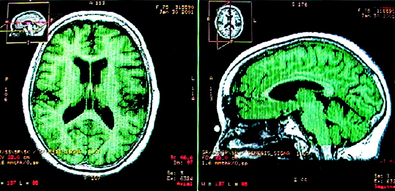
- Fig 1.
T1-weighted MR images display the relative global cerebral volume in a patient with AD in axial and sagittal views. Green represents the final region where the volume calculation was performed.
- Fig 2.
T1-weighted MR images display the relative hippocampal volume in a patient with AD in the axial and sagittal planes. Green represents the final region where the volume calculation was performed.
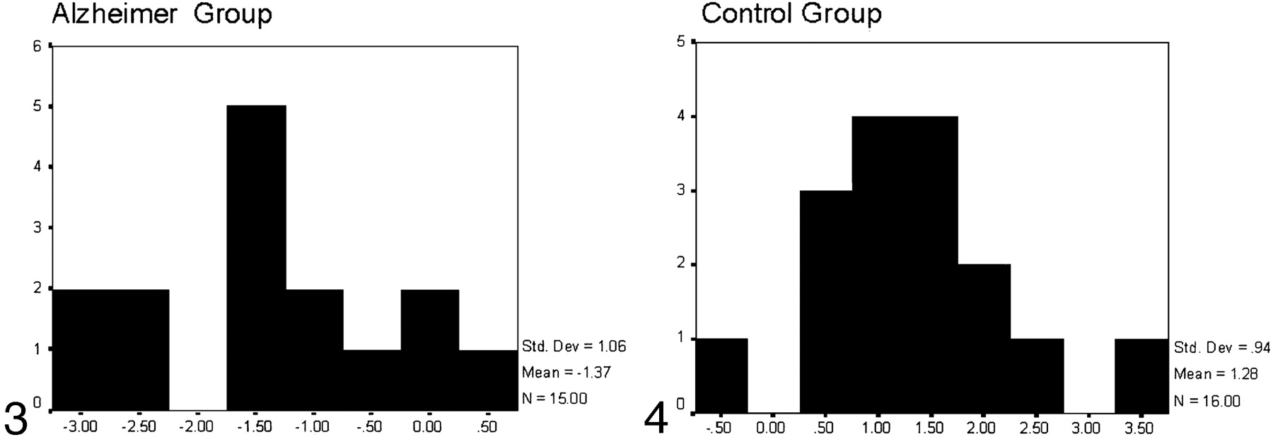
- Fig 3.
Histogram representing the results of the discriminant analysis for the Alzheimer group: x axis reflects canonical scores, and y axis reflects the number of subjects who obtained every canonical score. Those with canonical scores below zero were classified as belonging to the AD group; three controls were incorrectly classified within the AD group.
- Fig 4.
Histogram representing the results of the discriminant analysis for the control group: x axis reflects canonical scores, and y axis reflects the number of subjects who obtained every canonical score. Those with canonical scores greater than zero were classified as controls; one AD patient was incorrectly classified as belonging to the control group.


Tables
Measure and Group Mean SD Age, y AD 76.27 1.21 Control 73.68 1.32 CAMCOG AD 55.66 6.01 Control 80.89 7.83 MMSE AD 19.93 2.28 Control 31.36 2.49 FAST AD 3.46 0.5 Control 1.36 0.59 Note.—Regarding MMSE scores, in the Spanish version by Lobo and Ezquerra (26), the maximum score is 35.
Variable Delta Band Variable Theta Band Mean SD Mean SD A: AD group LF_D .406 .094 LF_T .868 .221 RF_D .327 .079 RF_T .811 .294 LP_D 1.190* .209 LP_T 1.959* .333 RP_D .982* .162 RP_T 2.418* .405 LPF_D .794 .174 LPF_T .682 .146 RPF_D .604 .175 RPF_T .410 .117 LT_D .313* .071 LT_T .531* .119 RT_D .225 .059 RT_T .833* .185 LO_D .219 .108 LO_T .320 .145 RO_D .256 .093 RO_T .328 .106 B: Control group LF_D .213 .091 LF_T .545 .214 RF_D .193 .076 RF_T .395 .284 LP_D .354 .202 LP_T .884 .322 RP_D .288 .157 RP_T .879 .392 LPF_D .435 .168 LPF_T .454 .141 RPF_D .459 .169 RPF_T .381 .113 LT_D 6.140E-02 .068 LT_T .141 .115 RT_D 6.284E-02 .057 RT_T .189 .180 LO_D 4.439E-02 .105 LO_T .225 .141 RO_D 7.432E-02 .090 RO_T .178 .103 Note.—Abbreviations are as follows: LF_D, left frontal delta; RF_D, right frontal delta; LP_D, left parietal delta; RP_D, right parietal delta; LPF_D, left prefrontal delta; RPF_D, right prefrontal delta; LT_D, left temporal delta; RT_D, right temporal delta; LO_D, left occipital delta; RO_D, right occipital delta; LF_T, left frontal theta; RF_T, right frontal theta; LP_T, left parietal theta; RP_T, right parietal theta; LPF_T, left prefrontal theta; RPF_T, right prefrontal theta; LT_T, left temporal theta; RT_T, right temporal theta; LO_T, left occipital theta; and RO_T, right occipital theta.
* Variables with significant differences between groups. For more information, see Fernández et al (22).
MEG Variable MRI Variable CVr RTVr LTVr RHVr LHVr Left parietal_delta −.35* −.23 −.35† −.42‡ −.47§ Right parietal_delta −.26 −.25 −.39† −.37† −.41‡ Left temporal_delta −.29 −.12 −.21 −.33* −.46§ Left parietal_theta −.38† −.25 −.37‡ −.40‡ −.44§ Right parietal_theta −.29 −.20 −.30 −.26 −.34 Left temporal_theta −.34* −.26 −.29 −.31* −.40‡ Right temporal_theta −.11 −.23 −.30 −.21 −0.22 * P < .05.
† P < .025.
‡ P < .01.
§ P < .002.
Group Predicted Group Total AD Control Original Count AD 12 3 15 Control 1 15 16 Ungrouped cases 3 5 8 Percentage, % AD 80.0 20.0 100.0 Control 6.3 93.8 100.0 Ungrouped cases 37.5 62.5 100.0 Cross-validated Count AD 12 3 15 Control 1 15 16 Percentage, % Alzheimer 80.0 20.0 100.0 Control 6.3 93.8 100.0


