Advertisement

Main menu

  • Home
  • Content
    • Current Issue
    • Accepted Manuscripts
    • Article Preview
    • Past Issue Archive
    • Video Articles
    • AJNR Case Collection
    • Case of the Week Archive
    • Case of the Month Archive
    • Classic Case Archive
  • Special Collections
    • AJNR Awards
    • Low-Field MRI
    • Alzheimer Disease
    • ASNR Foundation Special Collection
    • Photon-Counting CT
    • View All
  • Multimedia
    • AJNR Podcasts
    • AJNR SCANtastic
    • Trainee Corner
    • MRI Safety Corner
    • Imaging Protocols
  • For Authors
    • Submit a Manuscript
    • Submit a Video Article
    • Submit an eLetter to the Editor/Response
    • Manuscript Submission Guidelines
    • Statistical Tips
    • Fast Publishing of Accepted Manuscripts
    • Graphical Abstract Preparation
    • Imaging Protocol Submission
    • Author Policies
  • About Us
    • About AJNR
    • Editorial Board
    • Editorial Board Alumni
  • More
    • Become a Reviewer/Academy of Reviewers
    • Subscribers
    • Permissions
    • Alerts
    • Feedback
    • Advertisers
    • ASNR Home

User menu

  • Alerts
  • Log in

Search

  • Advanced search
American Journal of Neuroradiology

American Journal of Neuroradiology

ASHNR American Society of Functional Neuroradiology ASHNR American Society of Pediatric Neuroradiology ASSR
  • Alerts
  • Log in

Advanced Search

  • Home
  • Content
    • Current Issue
    • Accepted Manuscripts
    • Article Preview
    • Past Issue Archive
    • Video Articles
    • AJNR Case Collection
    • Case of the Week Archive
    • Case of the Month Archive
    • Classic Case Archive
  • Special Collections
    • AJNR Awards
    • Low-Field MRI
    • Alzheimer Disease
    • ASNR Foundation Special Collection
    • Photon-Counting CT
    • View All
  • Multimedia
    • AJNR Podcasts
    • AJNR SCANtastic
    • Trainee Corner
    • MRI Safety Corner
    • Imaging Protocols
  • For Authors
    • Submit a Manuscript
    • Submit a Video Article
    • Submit an eLetter to the Editor/Response
    • Manuscript Submission Guidelines
    • Statistical Tips
    • Fast Publishing of Accepted Manuscripts
    • Graphical Abstract Preparation
    • Imaging Protocol Submission
    • Author Policies
  • About Us
    • About AJNR
    • Editorial Board
    • Editorial Board Alumni
  • More
    • Become a Reviewer/Academy of Reviewers
    • Subscribers
    • Permissions
    • Alerts
    • Feedback
    • Advertisers
    • ASNR Home
  • Follow AJNR on Twitter
  • Visit AJNR on Facebook
  • Follow AJNR on Instagram
  • Join AJNR on LinkedIn
  • RSS Feeds

AJNR Awards, New Junior Editors, and more. Read the latest AJNR updates

Research ArticleHead and Neck Imaging

MR Imaging Study of Edema-like Change along the Optic Tract in Patients with Pituitary Region Tumors

Naokatsu Saeki, Yoshio Uchino, Hisayuki Murai, Motoo Kubota, Kouichi Isobe, Takashi Uno, Kenro Sunami and Akira Yamaura
American Journal of Neuroradiology March 2003, 24 (3) 336-342;
Naokatsu Saeki
  • Find this author on Google Scholar
  • Find this author on PubMed
  • Search for this author on this site
Yoshio Uchino
  • Find this author on Google Scholar
  • Find this author on PubMed
  • Search for this author on this site
Hisayuki Murai
  • Find this author on Google Scholar
  • Find this author on PubMed
  • Search for this author on this site
Motoo Kubota
  • Find this author on Google Scholar
  • Find this author on PubMed
  • Search for this author on this site
Kouichi Isobe
  • Find this author on Google Scholar
  • Find this author on PubMed
  • Search for this author on this site
Takashi Uno
  • Find this author on Google Scholar
  • Find this author on PubMed
  • Search for this author on this site
Kenro Sunami
  • Find this author on Google Scholar
  • Find this author on PubMed
  • Search for this author on this site
Akira Yamaura
  • Find this author on Google Scholar
  • Find this author on PubMed
  • Search for this author on this site
  • Article
  • Figures & Data
  • Info & Metrics
  • Responses
  • References
  • PDF
Loading

Article Figures & Data

Figures

  • Tables
  • Fig 1.
    • Download figure
    • Open in new tab
    • Download powerpoint
    Fig 1.

    Images from the case of a 59-year-old man with a giant pituitary adenoma.

    A, Midsagittal contrast-enhanced T1-weighted MR image obtained before treatment. A huge tumor (arrowhead) is visible in and on the sella turcica.

    B, Axial heavily T2-weighted MR image obtained one section above the level of the optic tract before treatment. Tumor (arrowhead) is visible in the third ventricle. Edema-like changes are visible bilaterally and are more prominent on the left side of the brain (right side of the figure) (large arrows). Large Virchow-Robin spaces related to anterior perforated substance are also visible (small arrows).

    C, Coronal heavily T2-weighted MR image obtained before treatment. Large Virchow-Robin space (large arrow) is located on the right optic tract (small arrow). Medium sized arrow and arrowhead indicate the edema-like change on the left side and the tumor, respectively.
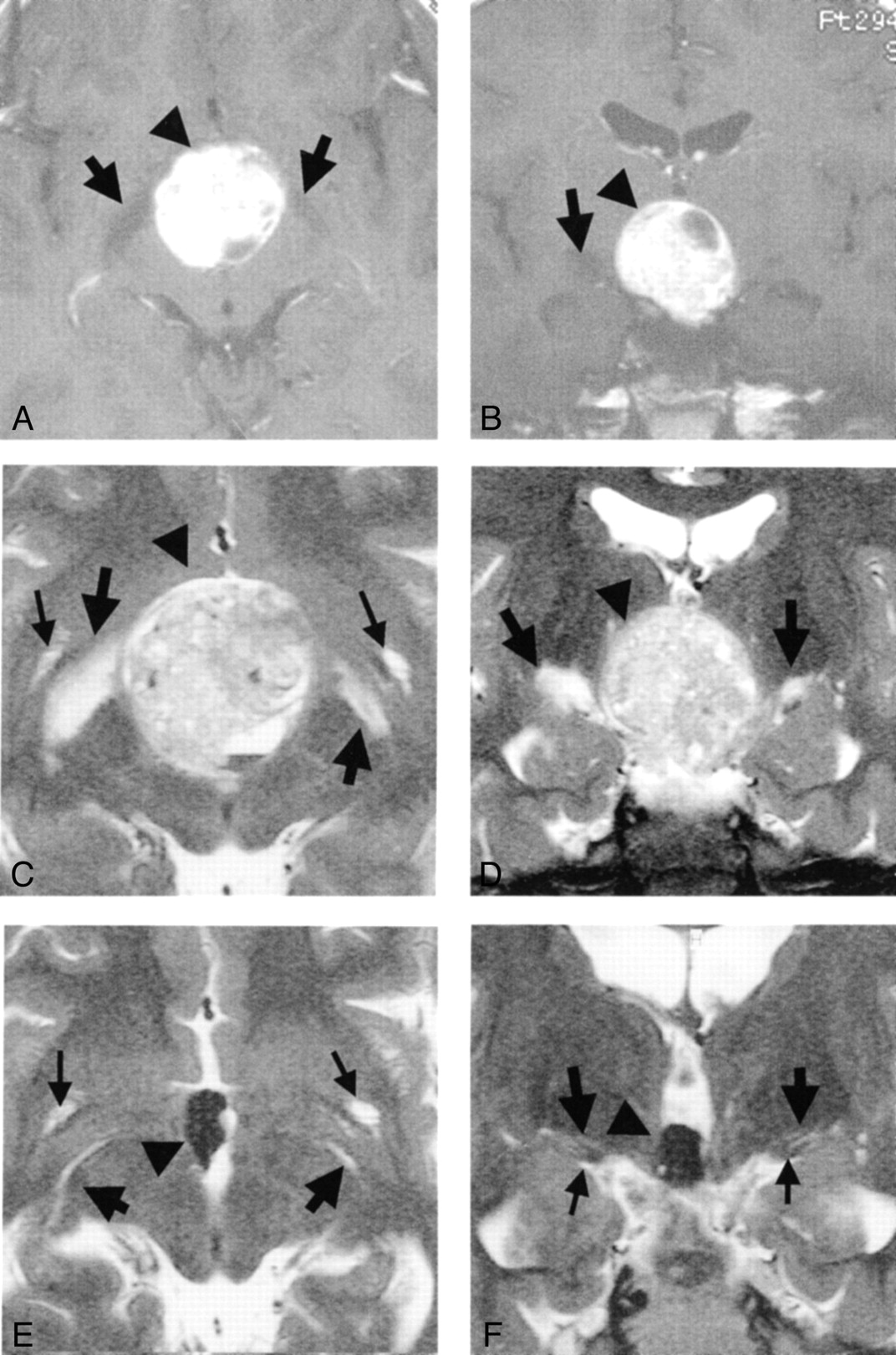
  • Fig 2.
    • Download figure
    • Open in new tab
    • Download powerpoint
    Fig 2.

    Images from the case of a 33-year-old woman with craniopharyngioma.

    A, Axial contrast-enhanced T1-weighted MR image obtained before treatment. Arrow and arrowhead indicate edema-like change and tumor, respectively.

    B, Coronal contrast-enhanced T1-weighted MR image obtained before treatment. High and low signal mass (arrowhead) is visible at the suprasellar cistern. Low signal edema-like change (arrow) is noted along the left optic tract.

    C, Axial heavily T2-weighted MR image obtained before treatment. Arrows indicate edema-like change.

    D, Coronal heavily T2-weighted MR image obtained before treatment. Edema-like changes (large arrows) are visible bilaterally along the optic tract and are more prominent on the left side of the brain (right side of the figure). Optic tracts are difficult to differentiate from edema-like changes. On the right side, a curvilinear high signal intensity (small arrow), originating from the edema-like change, is visible.

    E, Axial heavily T2-weighted MR image obtained one section above the level of the optic tract 3 months after surgery (at level similar to that shown in C). Edema-like change has disappeared on the right side of the brain. Large Virchow-Robin spaces (arrow, right side of brain), which are present under normal conditions, are visible on the same side. Edema-like change (arrow, left side of brain) seems to remain on the left side.

    F, Coronal heavily T2-weighted MR image obtained 3 months after surgery (at level similar to that shown in D). Large Virchow-Robin space (large arrow, right side of brain) is visible on the right optic tract (small arrow). Edema-like change (large arrow, left side of brain) remains on the left side.

  • Fig 3.
    • Download figure
    • Open in new tab
    • Download powerpoint
    Fig 3.

    Images from the case of an 11-year-old boy with a germ cell tumor.

    A, Axial contrast-enhanced T1-weighted MR image obtained before treatment. Edema-like change along the optic tract is visible bilaterally (arrows). Arrowhead indicates slightly hyperintense mass.

    B, Coronal contrast-enhanced T1-weighted MR image obtained before treatment. Round and almost high signal mass (arrowhead) is visible at the suprasellar region. Edema-like change along the optic tract is visible on the right side of the brain (left side of the figure) (arrow).

    C, Axial heavily T2-weighted MR image obtained before treatment. Large arrows, small arrows, and arrowheadindicate edema-like changes, Virchow-Robin spaces, and tumor, respectively. Findings concur with those in D.

    D, Coronal heavily T2-weighted MR image obtained before treatment. Edema-like changes (large arrows), more prominent on the right side of the brain (left side of the figure), are visible along the optic tracts. Tumor (arrowhead) is visible. Large Virchow-Robin spaces associated with anterior perforated substance are visible.

    E, Axial heavily T2-weighted MR image obtained 2 months after treatment (at level similar to that shown in C). Large Virchow-Robin spaces associated with anterior perforated substance (small arrows) are visible.

    F, Coronal heavily T2-weighted MR image obtained 2 months after treatment (at level similar to that shown in D). Edema-like change has disappeared bilaterally, although the tumor (arrowhead) remains in the third ventricle. Normally present large Virchow-Robin spaces (large arrows) are visible. The optic tracts (small arrows) are also visible. Large arrows and arrowhead indicate Virchow-Robin spaces and tumor, respectively.

  • Fig 4.
    • Download figure
    • Open in new tab
    • Download powerpoint
    Fig 4.

    Images from the case of a 62-year-old man with malignant lymphoma.

    A, Axial contrast-enhanced T1-weighted MR image obtained before treatment. Thick arrows, thin arrow, and arrowhead indicate edema-like change, incidentally found venous angioma, and tumor, respectively.

    B, Coronal contrast-enhanced T1-weighted MR image obtained before treatment. High and low signal mass (arrowhead) is visible in the third ventricle. Low signal edema-like changes (arrows) are noted along the optic tracts. A venous angioma was incidentally found in the left anterior basal ganglia, as in A.

    C, Axial heavily T2-weighted MR image obtained before treatment. Tumor (arrowhead) is visible. Marked edema-like changes are visible in the hypothalamus and midbrain and along the optic tracts (arrows).

    D, Coronal heavily T2-weighted MR image obtained before treatment. Tumor (arrowhead) is visible. Marked edema-like changes are visible in the hypothalamus and midbrain and along the optic tracts (arrows).

    E, Axial heavily T2-weighted MR image obtained before treatment. Edema-like changes (large arrows) extend as far as the basal ganglia and the internal capsule. The venous angioma (small arrow) is visible in the left anterior basal ganglia.

    F, Coronal heavily T2-weighted MR image obtained before treatment. Edema-like changes (large arrows) extend as far as the basal ganglia and the internal capsule. Marked edema-like change is visible in the hypothalamus and midbrain (small arrow).

Tables

  • Figures
    • View popup
    TABLE 1:

    Clinical profiles of 50 pituitary tumors

    HistologyNo. of PatientsSex (M:F)Age (yr)Edema-like Change Shown by MR Imaging (+:−)Tumor Size (Height) (mm)Visual Sign (+:−)
    Pituitary adenoma2511:1423–734:2121–5220:5
    Craniopharyngioma115:612–688:322–398:3
    Meningioma71:631–720:719–587:0
    Rathke’s cleft cyst53:234–630:519–275:0
    Germ cell tumor11:0111:0340:1
    Malignant lymphoma11:0621:0291:0
PreviousNext
Back to top

In this issue

American Journal of Neuroradiology: 24 (3)
American Journal of Neuroradiology
Vol. 24, Issue 3
1 Mar 2003
  • Table of Contents
  • Index by author
Advertisement
Print
Download PDF
Email Article

Thank you for your interest in spreading the word on American Journal of Neuroradiology.

NOTE: We only request your email address so that the person you are recommending the page to knows that you wanted them to see it, and that it is not junk mail. We do not capture any email address.

Enter multiple addresses on separate lines or separate them with commas.
MR Imaging Study of Edema-like Change along the Optic Tract in Patients with Pituitary Region Tumors
(Your Name) has sent you a message from American Journal of Neuroradiology
(Your Name) thought you would like to see the American Journal of Neuroradiology web site.
CAPTCHA
This question is for testing whether or not you are a human visitor and to prevent automated spam submissions.
Cite this article
Naokatsu Saeki, Yoshio Uchino, Hisayuki Murai, Motoo Kubota, Kouichi Isobe, Takashi Uno, Kenro Sunami, Akira Yamaura
MR Imaging Study of Edema-like Change along the Optic Tract in Patients with Pituitary Region Tumors
American Journal of Neuroradiology Mar 2003, 24 (3) 336-342;

Citation Manager Formats

  • BibTeX
  • Bookends
  • EasyBib
  • EndNote (tagged)
  • EndNote 8 (xml)
  • Medlars
  • Mendeley
  • Papers
  • RefWorks Tagged
  • Ref Manager
  • RIS
  • Zotero
0 Responses
Respond to this article
Share
Bookmark this article
MR Imaging Study of Edema-like Change along the Optic Tract in Patients with Pituitary Region Tumors
Naokatsu Saeki, Yoshio Uchino, Hisayuki Murai, Motoo Kubota, Kouichi Isobe, Takashi Uno, Kenro Sunami, Akira Yamaura
American Journal of Neuroradiology Mar 2003, 24 (3) 336-342;
del.icio.us logo Twitter logo Facebook logo Mendeley logo
  • Tweet Widget
  • Facebook Like
  • Google Plus One

Jump to section

  • Article
    • Abstract
    • Methods
    • Results
    • Discussion
    • Conclusion
    • Acknowledgments
    • References
  • Figures & Data
  • Info & Metrics
  • Responses
  • References
  • PDF

Related Articles

  • No related articles found.
  • PubMed
  • Google Scholar

Cited By...

  • Complete Regression of a Temporal Stem Dilated Perivascular Space Following Resection of a Pituitary Nonfunctioning Macroadenoma
  • Optic chiasm enhancement associated with giant aneurysm and yttrium treated pituitary adenoma
  • Crossref
  • Google Scholar

This article has not yet been cited by articles in journals that are participating in Crossref Cited-by Linking.

More in this TOC Section

  • Diagnostic Performance of US in Neck Node NIRADS 2
  • Hydrops Herniation into the Semicircular Canals
  • MRI of the Parasellar Ligaments
Show more HEAD AND NECK IMAGING

Similar Articles

Advertisement

Indexed Content

  • Current Issue
  • Accepted Manuscripts
  • Article Preview
  • Past Issues
  • Editorials
  • Editor's Choice
  • Fellows' Journal Club
  • Letters to the Editor
  • Video Articles

Cases

  • Case Collection
  • Archive - Case of the Week
  • Archive - Case of the Month
  • Archive - Classic Case

More from AJNR

  • Trainee Corner
  • Imaging Protocols
  • MRI Safety Corner
  • Book Reviews

Multimedia

  • AJNR Podcasts
  • AJNR Scantastics

Resources

  • Turnaround Time
  • Submit a Manuscript
  • Submit a Video Article
  • Submit an eLetter to the Editor/Response
  • Manuscript Submission Guidelines
  • Statistical Tips
  • Fast Publishing of Accepted Manuscripts
  • Graphical Abstract Preparation
  • Imaging Protocol Submission
  • Evidence-Based Medicine Level Guide
  • Publishing Checklists
  • Author Policies
  • Become a Reviewer/Academy of Reviewers
  • News and Updates

About Us

  • About AJNR
  • Editorial Board
  • Editorial Board Alumni
  • Alerts
  • Permissions
  • Not an AJNR Subscriber? Join Now
  • Advertise with Us
  • Librarian Resources
  • Feedback
  • Terms and Conditions
  • AJNR Editorial Board Alumni

American Society of Neuroradiology

  • Not an ASNR Member? Join Now

© 2025 by the American Society of Neuroradiology All rights, including for text and data mining, AI training, and similar technologies, are reserved.
Print ISSN: 0195-6108 Online ISSN: 1936-959X

Powered by HighWire