
Article Figures & Data
Figures
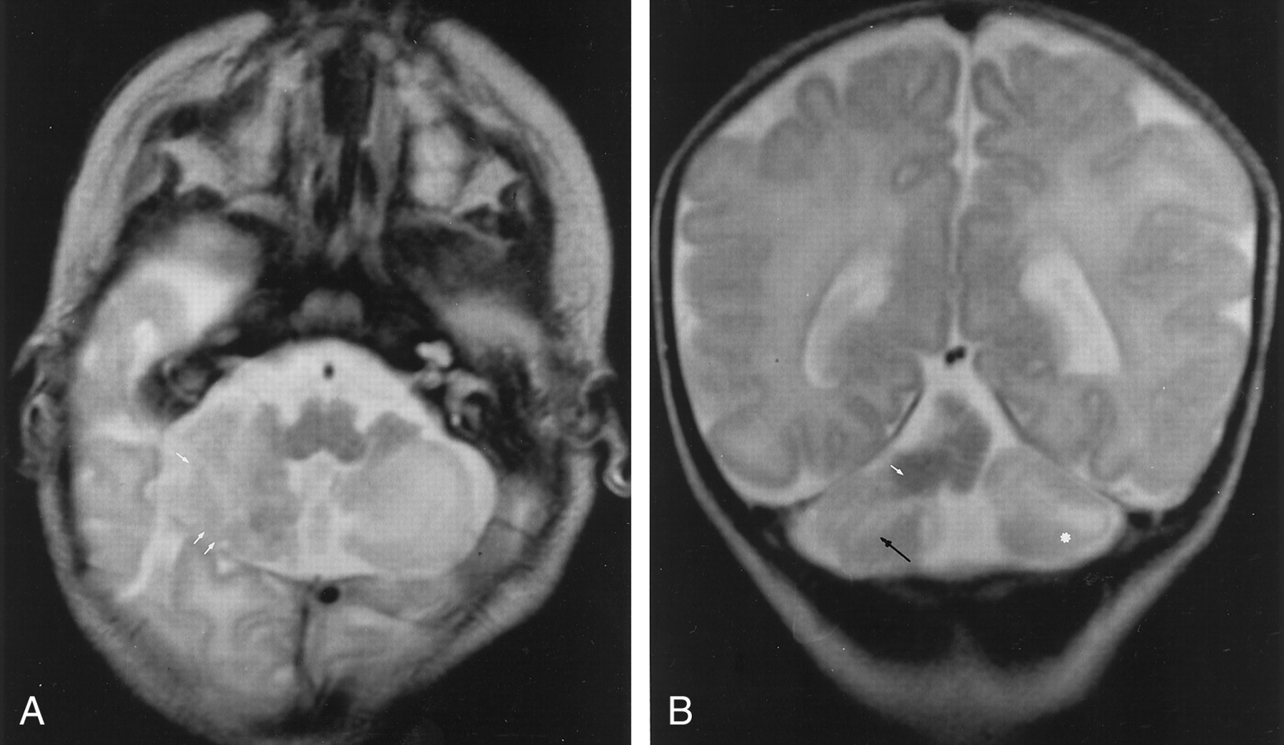
- Fig 1.
MR images in CCD associated with heterotopic and vermian hypoplasia.
A, Axial T2-weighted image (5000/12/2 [TR/TE/NEX]) shows vertically oriented and disorganized folia (white arrows) in the posteroinferior part of the hemisphere.
B, Coronal T2-weighted image (5000/120/2) shows vertically oriented fissures (black arrow) in the cerebellar cortex, consistent with the diagnosis of CCD. Note cerebellar white matter hyperintensity (white dot) and heterotopia (white arrow).
- Fig 2.
Coronal microscopic sections (posterior to anterior view) of the cerebellar lesions at vermian (top) and hemispheric (bottom) levels show vermian hypoplasia with irregularly shaped foliation. A large central multinodular conglomerate (arrows) corresponding to juxtaposed ectopic polymicrogeria nodules is seen in the right hemisphere and vermis.
- Fig 3.
Histologic sections in CCD associated with heterotopic and vermian hypoplasia.
A, Vermis and right cerebellar hemisphere show abnormal cerebellar lamella associated with typical polymicrogyria: deep folding of the cerebellar surface (black arrows). Note the heterotopic aspect of polymicrogyria in the white matter: cavities appear obliterated (white arrows). (Luxor blue stain, original magnification ×25.)
B, Right cerebellar hemisphere shows abnormally large cerebellar folia (as in premature cerebellum) (white arrows) associated with a large nodule of compact polymicrogyria with obliterated cavities and containing mixed cells (spindle, granular cells, and large neurons) (black arrows). (Luxol blue stain, original magnification ×50.)


