
Article Figures & Data
Figures
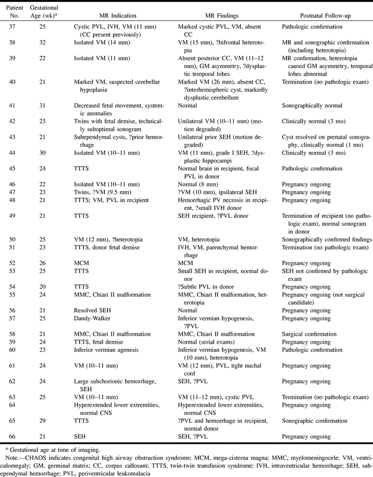
- fig 1.
Patient 13: isolated ventriculomegaly (23 weeks' gestation).
A and B, ssFSE T2-weighted images (∞/97/0.5) in the axial plane (A) show ventriculomegaly with 11-mm diameter at the atrium (arrow) and normal signal in the adjacent parenchyma. Coronal plane (B) reveals the normal hypointense signal of the germinal matrix (arrowheads).
- fig 2.
Patient 20: inferior vermian hypogenesis (23 weeks' gestation).
A, Axial ssFSE image (∞/98/0.5) shows the normal superior vermis (arrow).
B, Subjacent section shows CSF communication with the fourth ventricle (arrow).
C, Midline sagittal image shows hypogenetic inferior vermis with normal superior and hypoplastic inferior lobules (arrow).
- fig 3.
Patient 23: hydrolethalus syndrome with postnatal correlation (39 weeks' gestation).
A, Prenatal axial ssFSE image (∞/97/0.5) at the level of the third ventricle shows calvarial defect and meningocele (arrows).
B, Prenatal sagittal ssFSE image shows vermian hypogenesis, large fourth ventricle, elevated tentorium (Dandy-Walker malformation), and calvarial defects (arrows).
C and D, Postnatal axial (C) and sagittal (D) T2-weighted sequences (3000/120/1 and 3000/102/2, respectively) confirm the prenatal findings, although the meningoceles (arrows) are less apparent due to positional flattening.
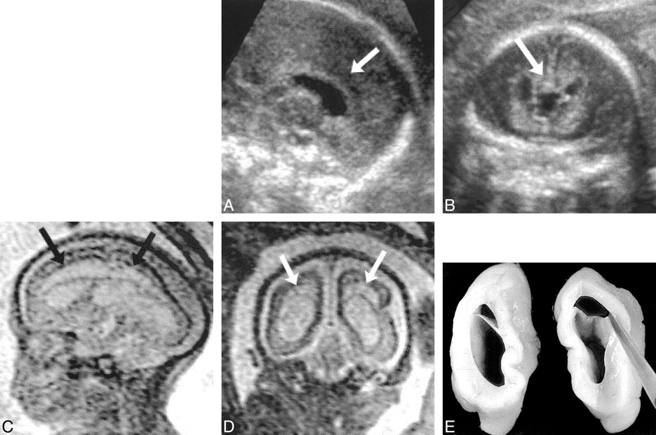
- fig 4.
Patient 37: cystic PVL and secondary absence of the corpus callosum (25 weeks' gestation).
A and B, Sagittal (A) and coronal (B) views from sonograms obtained at 22 weeks' gestation show normal corpus callosum (arrows).
C and D, Sagittal (C) and coronal (D) ssFSE images (∞/98/0.5) reveal development of cystic PVL (arrows) and absence of the corpus callosum.
E, Postmortem coronal section confirms the MR findings.
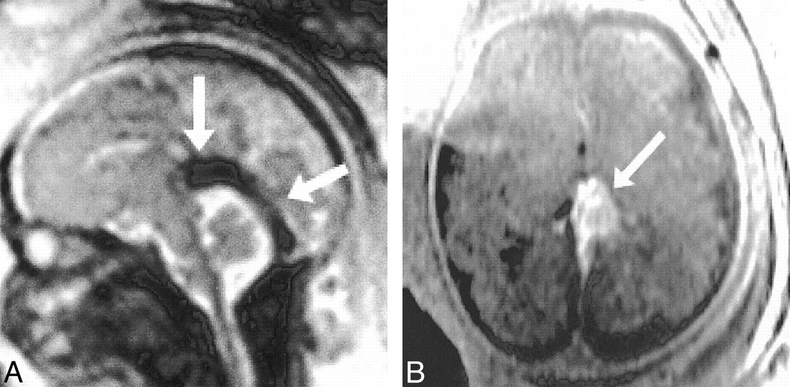
- fig 5.
Patient 39: hypogenesis of the corpus callosum (22 weeks' gestation).
A, Sagittal ssFSE image (∞/98/0.5) shows the genu and anterior body of the corpus callosum (arrow). The posterior body and splenium are absent.
B, Axial ssFSE image confirms the presence of the genu (white arrow). The cavum septi pellucidi (black arrows) is only seen when the genu and anterior callosal body are present. The absent splenium is apparent on this image, as the posterior interhemispheric fissure is continuous with the cavum.
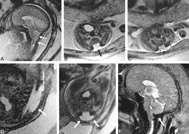
- fig 6.
Patient 17: Chiari II malformation and myelomeningocele (23 weeks' gestation).
A, Sagittal ssFSE image (∞/96/0.5) shows the poorly formed posterior fossa floor and downward cerebellar herniation (arrows).
B and C, Axial ssFSE images at the level of the lumbosacral region show absent posterior elements (arrow, B) and exposed neural elements (myelomeningocele, arrowhead, C).
D and E, Sagittal (D) and axial (E) ssFSE images (∞/98/0.5) 13 days after in utero repair show hypointense dural patch (arrows) over defect.
F, Sagittal ssFSE image (∞/97/0.5) approximately 10 weeks after repair suggests improved development of the floor of the posterior fossa (suboccipital bone, arrow) and reduced hindbrain herniation.
- fig 7.
Patient 30: vein of Galen malformation (33 weeks' gestation).
A, Sagittal ssFSE image (∞/98/0.5) shows large signal void caused by rapid flow in the vein of Galen varix and dilated straight sinus (arrows).
B, Axial FMPSPGR image (100/4.2/1) shows hyperintensity caused by flow-related enhancement in the varix (arrow).


Tables

Findings in 66 fetal MR examinations

In this issue


Jump to section
Related Articles
Cited By...
- Evaluation of Subependymal Gray Matter Heterotopias on Fetal MRI
- Complex Fetal Care: Implications of Fetal Ventriculomegaly: A Neurosurgical Perspective
- MRI of the foetal brain using a rapid 3D steady-state sequence
- The use of in utero MRI to supplement ultrasound in the foetus at high risk of developmental brain or spine abnormality
- The Use of In Utero MR Imaging to Delineate Developmental Brain Abnormalities in Multifetal Pregnancies
- High-Resolution In Utero 3D MR Imaging of Inner Ear Microstructures in Fetal Sheep
- Fetuses with Ventriculomegaly Diagnosed in the Second Trimester of Pregnancy by In Utero MR Imaging: What Happens in the Third Trimester?
- A Prospective Study of Fetuses with Isolated Ventriculomegaly Investigated by Antenatal Sonography and In Utero MR Imaging
- Fetal Magnetic Resonance Imaging in the Evaluation of Fetuses Referred for Sonographically Suspected Abnormalities of the Corpus Callosum
- Fetal Magnetic Resonance Imaging Enhances Detection of Spinal Cord Anomalies in Patients With Sonographically Detected Bony Anomalies of the Spine
- The Role of Magnetic Resonance Imaging in the Evaluation of Isolated Mild Ventriculomegaly